Examining MDMX / MDM2 Signaling Pathways in Breast Cancers Expressing Mutant p53
| Name | Rusia H. Lee |
| Institution | Hunter College, City University of New York |
| Research Field | Basic Research |
| Role at Institution | Graduate Student |
| Presenter(s) | Rusia H. Lee |
Examining MDMX / MDM2 Signaling Pathways in Breast Cancers Expressing Mutant p53
Rusia Lee1,2,3, Viola Ellison2, Dominique Forbes2, Gu Xiao2, Alexandra Kern2, Pam Leybengrub2, Jill Bargonetti1,2,3
1The Graduate Center- City University of New York (CUNY)
2The Department of Biological Sciences Hunter College
3Weill Cornell Medical College, New York City
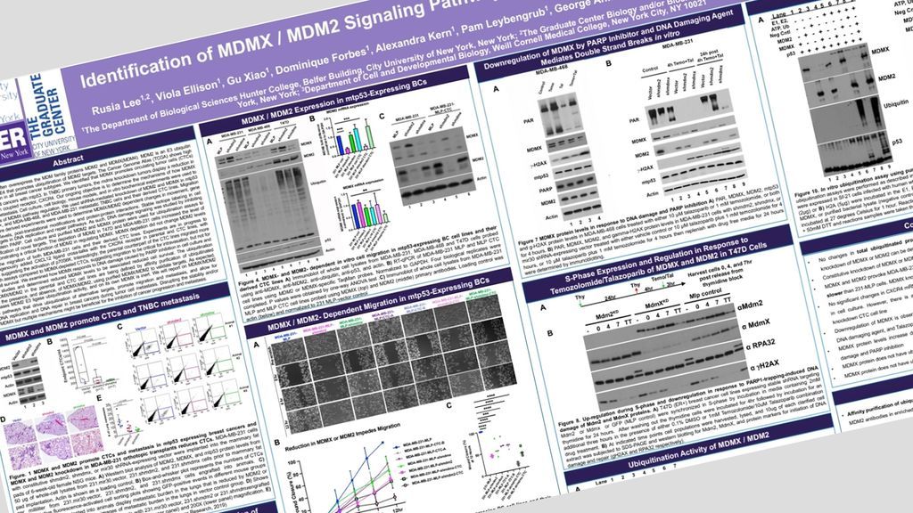
Mouse Double Minute 2 (MDM2) and Mouse Double Minute 4/X (MDM4 also called MDMX) are negative regulators of tumor suppressor protein p53. MDM2 is amplified in approximately ~10% of human cancers, and MDMX is amplified in 10-12% of breast cancers. MDMX forms heterodimer complexes with MDM2 to enhance MDM2 E3 ligase activity for wtp53. Importantly MDM2 and MDMX are often found over-expressed in the context mutant p53. However, the MDMX/MDM2 functions that are independent of targeting wtp53 for degradation remain unclear. There is a need to further study MDM2/MDMX wtp53-independent tumor promoting activities.
Using xenograft mouse models, we demonstrated that the MDMX/ MDM2 heterodimeric relationship in the presence of GOF mtp53 R280K plays a role in driving TNBC increased circulating tumor cell (CTC) formation and early metastasis. This pointed to MDM2/MDMX metastasis promotion working through a p53-independent pathway. We identified that MDMX knockdown in primary tumors correlates with a downregulation in CXCR4 and PTGS2 transcripts. Studies show that silencing CXCR4 in BC mouse models reduces metastatic burden,
TNBC cell lines, MDA-MB-231-MLP-vector control, MDA-MB-231-MLP-shmdm2, and MDA-MB-231-MLP-shmdmx, were orthotopically injected into xenograft mouse models. TNBC-derived CTC cell lines were established by growing CTCs in culture. We performed an in vitro migration assay to model the metastatic phenotype ex vivo using the MDA-MB-231-MLP and MDA-MB-231-MLP-CTC cells and their knockdown lines. We observed delayed migration with a reduction in MDMX or MDM2 in MDA-MB-231-MLP-CTC lines compared to 231-MLP cells. This suggests that MDMX or MDM2 induces cell migration in vitro in non-CTC (231-MLP) and CTC lines. Through RT-qPCR and immunoblot analysis, we observed a significant reduction in CXCR4 levels, which suggests that the MDMX-mediated early metastasis pathways occur prior to CTC formation.
To identify ubiquitin targets of the MDMX/MDM2 heterodimer complex, we enriched for ubiquitinated proteins through affinity purification. We will report on our preliminary data in this protein-target identification area, with a focus on CXCR4 and histones. Our objective is to identify novel MDMX or MDM2/MDMX heterodimer protein interactions that assist MDMX or MDM2 in driving TNBC metastasis independent of p53.
(n=338)