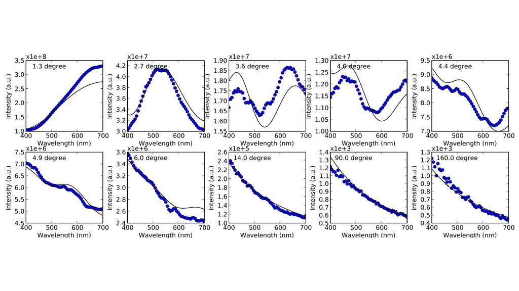
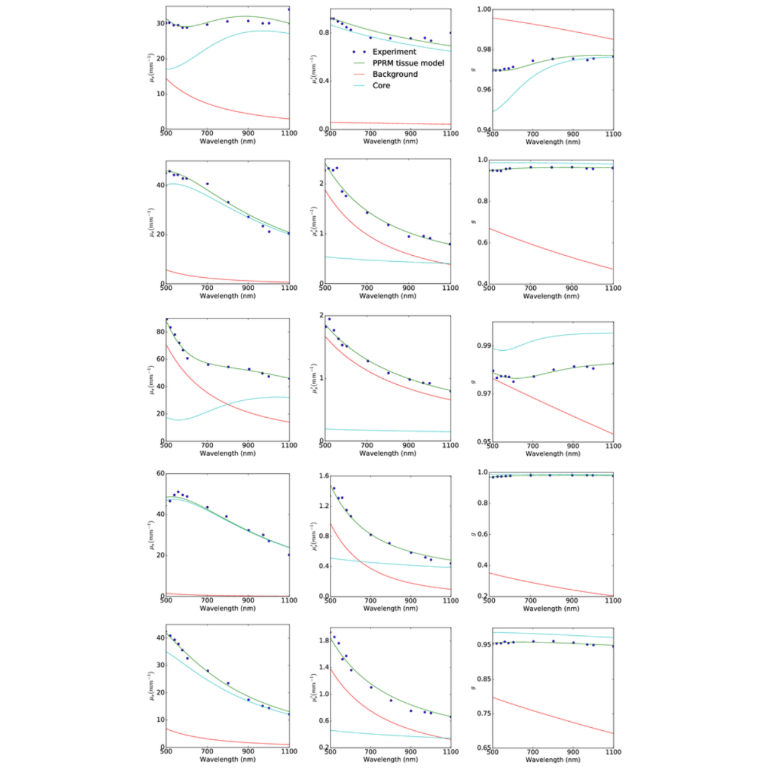
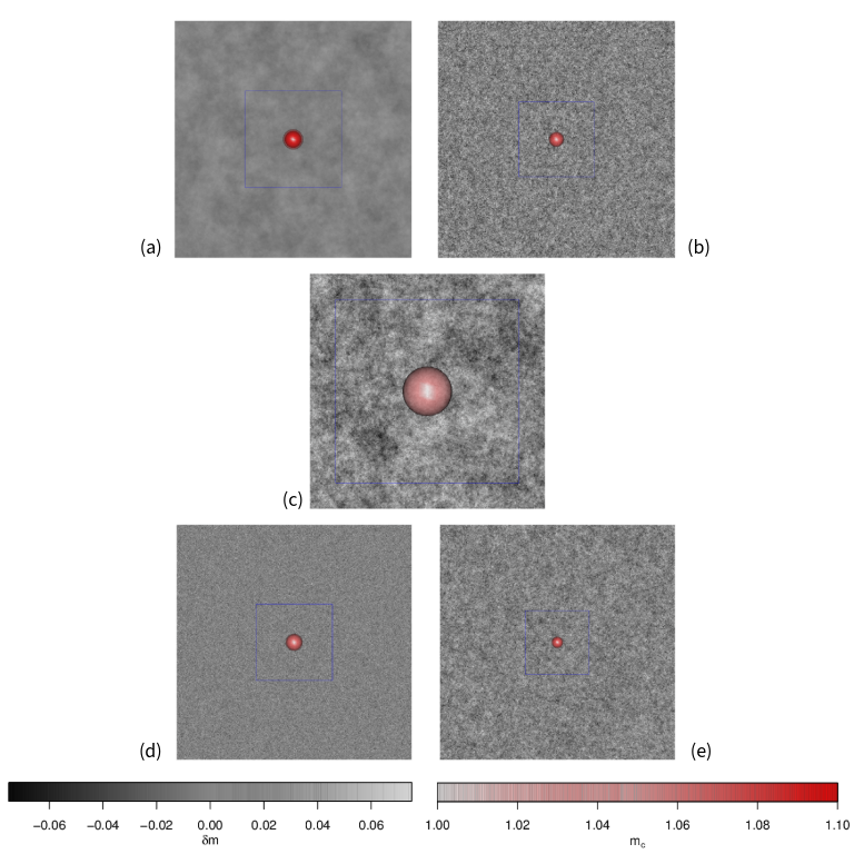
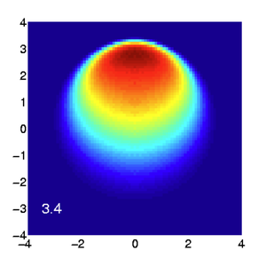
Hunter College
  • Information for
    • Students
    • Alumni & Friends
    • Faculty
    • Staff
    • Community
  • QUICK LINKS
  • DIRECTORY
  • APPLY
  • GIVE
  • RENT
Hunter College The School of Arts & Sciences
The School
  • Office of the Dean
  • Departments & Programs
  • Undergraduate Academics
  • Graduate Academics
  • Awards & Scholarships
  • Resources for Students
  • Resources for Faculty & Staff
  • Faculty Awards & Achievements
  • News & Events
  • Contact Us
Departments & Programs (A-H)
  • Africana, Puerto Rican & Latino Studies
  • Anthropology
  • Art & Art History
  • Asian American Studies
  • Biological Sciences
  • Chemistry
  • Classical & Oriental Studies
  • Computer Science
  • Creative Writing
  • Dance
  • Economics/Accounting
  • English
  • Film & Media Studies
  • Geography & Environmental Science
  • German
  • History
  • Human Biology
  • Human Rights
Departments & Programs (I-Z)
  • Jewish Studies
  • Latin American & Caribbean Studies
  • Macaulay Honors College
  • Mathematics & Statistics
  • Medical Laboratory Sciences
  • Music
  • Philosophy
  • Physics & Astronomy
  • Political Science
  • Psychology
  • Public Policy
  • Religion
  • Romance Languages
  • Sociology
  • Theatre
  • Thomas Hunter Honors Program
  • Translation & Interpreting
  • Urban Policy and Planning
  • Women & Gender Studies
  • Admissions
  • Advising
  • Libraries
  • Undergraduate Catalog
  • Graduate Catalog
 
Hunter College Home
Hunter College Schools
  • School of Arts & Sciences
  • School of Education
  • School of Health Professions
  • Hunter-Bellevue School of Nursing
  • Silberman School of Social Work
More Schools
  • Hunter College Campus Schools
  • Hunter College Continuing Education
  • Libraries
  • Undergraduate Catalog
  • Graduate Catalog
  • Students
  • Alumni & Friends
  • Faculty
  • Staff
  • Community
  • Admissions
  • Advising
  • APPLY
  • GIVE
  • RENT
  • QUICK LINKS
  • DIRECTORY
School of Arts and Sciences /
Physics and Astronomy
  • About
  • Undergraduate
  • Graduate
  • Courses
  • Faculty and Staff
  • Research
    • Biomedical Photonics Laboratory
    • Ultrafast Optics and Spectroscopy Lab
    • Laser Laboratory
    • Solid State NMR/EPR Laboratory
    • Staff
  • Resources
  • News
  • Contact

Biomedical Photonics Laboratory

Welcome

Lighting up static structure and dynamic processes in biological systems for biomedical applications.

Professors in lab

Overview

The quest for the physics of light-turbid soft matter (e.g., biological tissue and cells) interaction and the endeavor in engineering for noninvasively characterization and diagnosis of tissue and cells with light at the interface of physics, engineering and biomedical sciences are the current central themes of Biomedical Photonics Laboratory @ Hunter. With the recent advances in light sources, manipulation devices, and detectors as well as the ever-increasing computational power, biophotonics has attracted significant interest and is well positioned to tackle unmet needs and challenges in health-related imaging, monitoring and diagnosis. Biomedical Photonics Laboratory is devoted to address fundamental problems and make innovations in this exciting field with integrated analytical, computational, and experimental approaches.

Our Work

In one part, we have developed realistic light scattering models for biological tissue and cells, and worked on improving the modeling of light interaction with biological tissue and cells—depolarization, decoherence and propagation of multiply scattered light through biological systems—to establish a solid foundation for applying light to medicine. In the other part, we study the inverse problems arisen in applying light to characterize and diagnose biological tissue and cells, and develop novel approaches for biomedical optical spectroscopy and imaging.

Our current research focuses on:

  1. Using light to noninvasively probe the physiological and functional change of biological systems and detect tumors at the early stage of development in both subsurface and deep tissues with engineered light;
  2. Development of label-free optical pathology for cancer diagnostics and prognostics with pathological specimens using quantitative microscopy and deep learning;
  3. Innovations in deep tissue and brain imaging and monitoring with interferometric diffuse optical imaging.

Research Projects

Quantitative modeling of tissue scattering and absorption properties is fundamental to determine light transport and energy deposition in tissue, key to both diagnostic and therapeutic applications of light. The rich spectroscopic behavior of biological tissue arising from its complex structure, as one main strength of optical interaction with biological tissue and cells compared to other biomedical imaging modalities, has also provided a powerful noninvasive probe to structural alterations in tissue due to disease or physiology. It is hence critical to establish connection between the characteristics of light scattering to the complex structure of tissue and cells.

A fractal continuous random medium model [1, 2, 3] has been proposed for light scattering by biological medium based on the observation that many biological tissues have fractal-like organization and on a macroscopic scale the constituents of tissue have no clear boundaries and merge into a quasi-continuum structure. The model has been shown to describe well light scattering by both tissue and cell suspensions. Via a unified Mie and fractal model derived from the superposition rule for light scattering by a composite particle [4], the fractal dimension and the correlation length reflecting both the morphological structure of tissue and the nuclear structure can be detected by spectroscopic light scattering measurement [3, 5]. The unified model is currently the only analytical model for light scattering by cells that describes correctly both the Mie resonance structure near forward scattering direction and the fractal scattering at larger angles. Preliminary experimental results show the fractal model can be successfully applied to discriminate accurately normal and malignant prostate tissues [6].


research-Scattering0x

Figure 1: Experimental (solid circles) and unified model fitting (solid line) results for the scattering spectra of the SiHa cell suspension at 10 representative scattering angles. The size distributions for the cell and the nucleus were measured separately by microscopy and not fitted. Note the Mie resonance at small scattering angles and the fractal scattering behavior at larger angles.


A novel statistical theory of light extinction by soft particles [7, 8] has been developed and successfully applied to characterize bacteria and other soft particles of arbitrary shapes and sizes in vivo [9, 10]. For example, both refractive index and size change of Bacillus subtilis endospores during heat-shock-induced activation have been monitored in situ [10]. This technique has applications in biosensing of bacterias. Light scattering by fractal aggregates has also been studied to monitor the aggregation of clays [11].

The latest line of research is to characterize the superficial layer of tissue where approximately 90% of cancer initiates using low coherence enhanced backscattering of light [12, 13]. Enhanced backscattering is one fascinating physical phenomenon (also known as weak localization) of multiple scattering light that originates from the constructive interference between the light waves propagating along a pair of time-reversal trajectories. Low spatial coherence of incident light ensures interference only occurs for light remitting within the coherence length away from the incident point and hence limits the penetration depth of low coherence enhanced backscattering light. The preliminary results show that the low coherence enhanced backscattering spectrum can be used to extract both the dominant scatterer size and the fractal dimension of the medium. Being able to probe the nuclear structural change noninvasively at subsurface is significant and finds numerous applications in cancer screening and diagnostics.

Plum pudding random medium model for biological tissue 

Variations in the refractive index cause tissue to scatter light. Light of longer wavelength is expected to be more isotropically scattered into all directions as the scatterers appear smaller with respect to the wavelength and the anisotropy factor (g) defined as the mean cosine of the scattering angles gets smaller. This widely-accepted wavelength dependence of g is, however, contradictory to that found by thorough measurements within visible and near-infrared spectral range. The "anomalous" increase of g with the probing wavelength seems to be the rule rather than the exception for tissue light scattering. The anisotropy factor is one central parameter in photon multiple scattering and migration that bears the direct relation to the morphology and optical properties of the underlying microscopic scattering constituents. This contradiction between experiments and theory on g, in particular, reveals the current lack in the understanding of the nature of tissue light scattering.

We have developed, for the first time, a plum pudding random medium (PPRM) model [14] for biological tissue. PPRM properly describes tissue as a continuum of background refractive index fluctuation (pudding) yet with some prominent structures (plum) which are distinctive from the background medium. In this unified view, tissue light scattering is a superposition of both plums and pudding [4, 3]. The former includes, for example, the nuclear structure in soft tissue and fiber bundles in muscle. The latter incorporates smaller scattering structures such as organelles and refractive index variations throughout the tissue continuum. The powerlaw dependence of the reduced scattering coefficient is attributed to the fractal scattering pudding [1] whereas the distinctive prominent structure is responsible for the observed "anomalous" anisotropy trend. PPRM provides a potential resolution to the long-lasting puzzle in the spectroscopic properties of tissue.


Figure 2: Plum pudding random medium tissue model fitting to from top row to bottom row (a) normal breast adipose tissue, (b) normal glandular breast tissue, (c) fibrocystic tissue, (d) fibroadenoma, and (e) ductal carcinoma. The columns from left to right show the scattering coefficient μs, the reduced scattering coefficient μs′, and the anisotropy factor g. The background refractive index fluctuation and the core are shown together with the PPRM tissue model. Experimental data is adapted from Peters et al, Phys. Med. Biol., 35(9):1317-1334, 1990.


In vivo optical histopathology of deep tissue has long been one coveted goal of biophotonics. PPRM not only offers an analytical platform to understand and interpret light scattering by the complex structures in tissue but also have opened up a novel venue of quantifying the tissue architecture and microscopic structures from macroscopic probing of the bulk with scattered light alone. The determination of the background refractive index fluctuation and the properties of the plums completely characterizes and can further depict the tissue architecture and microscopic structure on average. PPRM may establish the foundation towards achieving such extremely desirable remote microscopy of biological tissue derived from non-contact spectroscopic light scattering measurement without tissue excision. This potential was demonstrated by visualizing the fine microscopic structural alterations in breast tissue (adipose, glandular, fibrocystic, fibroadenoma, and ductal carcinoma) deduced from noncontact spectroscopic measurement alone.


research-scattering3

Figure 3: The plum and pudding (background refractive index fluctuation) in (a) normal breast adipose tissue, (b) normal glandular breast tissue, (c) fibrocystic tissue, (d) fibroadenoma, and (e) ductal carcinoma derived from scattering spectroscopy measurement. The whole window size is 30μm × 30μm. The blue square delineates a unit cell which contains exactly one core. A core of most probable radius is shown, surrounded by a shaded area of radius at which the number density of the core drops to half maximum.


On-going research projects:

  • Deep tissue histopathology without tissue excision (remote microscopy) from spectroscopic light scattering based on the plum pudding random model of biological tissue.

Selected Publications

  1. M. Xu and R. R. Alfano. Fractal mechanisms of light scattering in biological tissue and cells. Opt. Lett., 30:3051-3053, 2005.
  2. Tao T. Wu, Jianan Y. Qu, and Min Xu. Unified Mie and fractal scattering by biological cells and subcellular structures. Opt. Lett., 32:2324-2326, 2007.
  3. M. Xu, Tao T. Wu, and Jianan Y. Qu. Unified Mie and fractal scattering by cells and experimental study on application in optical characterization of cellular and subcellular structures. J. Biomed. Opt., 13:038802, 2008.
  4. M. Xu. Superposition rule for light scattering by a composite particle. Opt. Lett., 31:3223-3225, 2006.
  5. Min Xu. Quantifying microarchitectural and light scattering differences between tumorigenic and non-tumorigenic cell models of tissue: analysis with unified Mie and fractal model. In Biomedical Optics/Digital Holography and Three-Dimensional Imaging/Laser Applications to Chemical, Security and Environmental Analysis on CD-ROM, page BTuF11, Optical Society of America, Washington, DC, 2008.
  6. Yang Pu, Wubao Wang, Mohammad AL-Rubaiee, Swapan Kumar Gayen, and Min Xu. Determination of optical coefficients and fractal dimensional parameters of cancerous and normal prostate tissues. Appl. Spectroscopy, 66:828-834, 2012.
  7. M. Xu, M. Lax, and R. R. Alfano. Light anomalous diffraction using geometrical path statistics of rays and gaussian ray approximation. Opt. Lett, 28:179-181, 2003.
  8. M. Xu and A. Katz. Light Scattering Reviews, volume III, chapter Statistical Interpretation of Light Anomalous Diffraction by Small Particles and its Applications in Bio-agent Detection and Monitoring, pages 27-68. Springer, 2008.
  9. A. Katz, A. Alimova, M. Xu, E. Rudolph, M. Shah, H. Savage, R. Rosen, S. A. McCormick, and R. R. Alfano. Bacteria size determination by elastic light scattering. IEEE JSTQE, 9:277-287, 2003.
  10. A. Katz, Alexandra Alimova, M. Xu, Paul Gottlieb, Elizabeth Rudolph, J. C. Steiner, and R. R. Alfano. In Situ determination of refractive index and size of Bacillus spores by light extinction. Opt. Lett., 30:589-591, 2005.
  11. Alexandra Alimova, A. Katz, Julian Orozco, Hui Wei, Paul Gottlieb, Elizabeth Rudolph, J. C. Steiner, and Min Xu. Broadband light scattering measurements of the time evolution of the fractal dimension of smectite clay aggregates. J. Opt. A, 11:105706, 2009. (Feature Article).
  12. Min Xu. Low coherence enhanced backscattering beyond diffusion. Opt. Lett., 33:1246-1248, 2008.
  13. Min Xu. Coherent backscattering of polarized light for tissue diagnostics: an electric field Monte Carlo study. In Steven L. Jacques, William P. Roach, and Robert J. Thomas, editors, Optical Interactions with Tissue and Cells XVIIII, 6854, page 68541A, 2008.
  14. Min Xu. Plum pudding random medium model of biological tissue toward remote microscopy from spectroscopic light scattering. Biomed. Opt. Express, 8:2879-2895, 2017.

Light interaction with random media is ubiquitous in nature and has found vast applications in applied sciences. Well-known examples for random media are biological tissue, atmosphere, ocean, and colloidal systems. The emergent light after interaction with a random medium (transillumination or backscattering) has been actively pursued as a means of non-invasively probing the internal structure of the random medium in the past decades (photonic diagnostics of random media).

The propagation of multiply scattered light in random media follows a universal law and light diffuses after a sufficient number of scattering events has occurred. Light diffusion is characterized by one length scale—the transport mean free path, lt, i.e., the distance over which the initial light propagation direction is randomized. By treating analytically the propagation of polarized light in turbid media as a random walk of vector photons, I have identified two additional length scales governing depolarization of the multiply scattering polarized light: (1) the characteristic length, lp, for polarized light to become isotropic in its linear polarization and propagation directions [1, 2], and (2) the characteristic length, lx, for circular polarized light to lose its helicity [3]. This also leads to a simple analytical model for the circular polarization memory effect of light. Other highlights of my relevant past work include a cumulant transport model for radiative transfer, providing a much more accurate transport model than the commonly used diffusion approximation [4, 5, 6]; an Electric Field Monte Carlo method (EMC) to simulate propagation, decoherence and depolarization of polarized light in random media; and low coherence enhanced backscattering of light [7, 8].


Propagation of a collimated beam in random media

Figure 1: Propagation of a collimated beam in random media. The x,y coordinates are measured in, lt, the transport mean free path.


On-going research projects:

  • Unification of light depolarization and decoherence into the model describing light propagation in random media. This work shall explore wave-shaping in the control of light propagation in turbid media and address the important practical problem where light is used to interrogate small turbid volumes or subsurface.

Selected Publications

  1. M. Xu, W. Cai, M. Lax, and R. R. Alfano. Stochastic view of photon migration in turbid media. arXiv:cond-mat/0401409, 2004.
  2. M. Xu and R. R. Alfano. Random walk of polarized light in turbid media. Phys. Rev. Lett., 95:213905, 2005.
  3. M. Xu and R. R. Alfano. Circular polarization memory of light. Phys. Rev. E, 72:065601(R), 2005.
  4. M. Xu, W. Cai, M. Lax, and R. R. Alfano. A photon transport forward model for imaging in turbid media. Opt. Lett., 26(14):1066-1068, 2001.
  5. M. Xu, W. Cai, M. Lax, and R. R. Alfano. Photon migration in turbid media using a cumulant approximation to radiative transfer. Phys. Rev. E, 65:066609, 2002.
  6. W. Cai, M. Xu, and R. R. Alfano. Analytical form of the particle distribution based on the cumulant solution of the elastic Boltzmann transport equation. Phys. Rev. E, 71:041202, 2005. (10 pages).
  7. Min Xu. Low coherence enhanced backscattering beyond diffusion. Opt. Lett., 33:1246-1248, 2008.
  8. Xiuwei Zhu, Luyao Lu, Zili Cao, Bixin Zeng, and Min Xu. Transmission matrix-based Electric field Monte Carlo study and experimental validation of the propagation characteristics of Bessel beams in turbid media. Opt. Lett., 43(19):4835, oct 2018.

Light multiple scattering is commonly studied using Monte Carlo simulations. The deficiency of conventional Monte Carlo methods is their lack of information about the phase of light and is not suitable for the study of coherence phenomena. A novel electric field Monte Carlo (EMC) method for simulation polarized light propagation in turbid media has recently been developed by us [1, 2]. The EMC method is different from conventional Monte Carlo approaches. EMC simulates the propagation of the electric field directly and hence contains the complete phase of light when it propagates in a turbid medium, accrued from both scattering by particles and propagation in the medium. This makes EMC a unique Monte Carlo approach able to study phase correlation and coherence effects of multiple scattering light in a first principle way. One example is the EMC study of coherent backscattering of polarized coherent light by a turbid medium [3].


Backscattering Mueller matrix

Figure 1: Backscattering Mueller matrix. All 4 × 4 matrix element are displayed as a two-dimensional image of the surface, 20ls × 20ls in size, with the laser being incident in the center and ls being the scattering length. The displayed Mueller matrix has been normalized by the maximum light intensity of the (1, 1) element.


Recently transmission matrix-based Electric field Monte Carlo (TEMC) method [4] has been introduced to study the propagation characteristics of Bessel beams with different orbital angular momentum (OAM) in turbid media. As an extension to the Electric field Monte Carlo (EMC) approach, electric field transmission modes were simulated to properly evaluate light interference. Experiment results agreed well with the theoretical simulations, validating the proposed TEMC for the study of coherence phenomenon in turbid media.


Electric field Monte Carlo

Figure 2: The comparison between experimental (2 and 4 rows) and TEMC simulation (1 and 3 rows) results of Bessel beams with OAM=1 and 5 at different thicknesses of z∕lt of 0, 1, 3 and 5.


Selected Publications

  1.  M. Xu. Electric field Monte Carlo for polarized light propagation in turbid media. Opt. Express, 12:6530-6539, 2004. http://www.opticsexpress.org/abstract.cfm?URI=OPEX-12-26-6530.
  2. Kevin G. Phillips, Min Xu, S. K. Gayen, and R. R. Alfano. Time-resolved ring structure of circularly polarized beams backscattered from forward scattering media. Opt. Express, 13:7954-7969, 2005.
  3. John Sawicki, Nikolas Kastor, and Min Xu. Electric field Monte Carlo simulation of coherent backscattering of polarized light by a turbid medium. Opt. Express, 16:5728-5738, 2008.
  4. Xiuwei Zhu, Luyao Lu, Zili Cao, Bixin Zeng, and Min Xu. Transmission matrix-based Electric field Monte Carlo study and experimental validation of the propagation characteristics of Bessel beams in turbid media. Opt. Lett., 43(19):4835, oct 2018.

Inverse problems in physical and biological sciences and biomedical imaging

Near infrared (NIR) light provides characterization and diagnostics of biological tissue and cells which is noninvasive, safe, of low cost, and easy to use. NIR light can penetrate deep (~5cm) into tissue. By probing the tissue with NIR light at multiple wavelengths, the concentration of the chromophores (oxy- and deoxy-hemoglobins, water and lipid) of tissue can be reconstructed from light attenuation within the tissue because the spectra of NIR light absorption by different chromophores differ significantly. The physiological and functional information of tissue can then be visualized in a three-dimensional (3D) image. The structural change caused by carcinogenesis results in the change of scattering property of tissue and can also be probed by light. With the current development in molecular beacons and other advanced fluorescence agents, light can detect tissue changes at the molecular level. However, strong scattering of light by tissue obscures direct images and inverse reconstruction is required to form images.

Inverse problem is ubiquitous in physical and biological sciences where unobservables are deduced from observable quantities. Moreover, inverse problem is inevitable in imaging a heterogeneous medium. In biomedical optical imaging where breast, prostate, or brain and etc. is being examined, challenges exist in both modeling accurately light propagation in biological tissue and in dealing with the ill-posedness of the inverse problem.

My research on optical imaging consists of developing a more accurate forward model than the commonly used diffusion approximation and advanced reconstruction methods. The cumulant solution to radiative transfer improved upon the diffusion approximation in optical imaging [1, 2, 3]. In the area of inverse reconstruction, several fast three-dimensional image reconstruction algorithms have been developed based on symmetry considerations and Fourier techniques [4, 5, 6], and recently a new scheme for optical imaging using independent component analysis (OPTICA) has been introduced [7, 8, 9, 10].

OPTICA interprets optical imaging as a source separation problem and uses independent component analysis to sort out the inhomogeneities within the turbid medium. OPTICA is fast and has several salient features compared to conventional optical imaging methods. In particular, a prior assumption about how light propagates in the medium is not needed to find out the inhomogeneities. A model for light propagation is only required in a Green’s function analysis of the retrieved independent components to locate and characterize the inhomogeneities. Unlike the conventional optical imaging methods, OPTICA doesn’t require an accurate modeling of light migration across short distances. Both simulations [8] and experiments [7, 9, 10] have shown OPTICA works best for small absorption, scattering, or fluorescence inhomogeneities (~ 1 - 10mm in size) and resolves the position and optical properties of localized inhomogeneities to a high degree of accuracy. This approach works extremely well for fluorescence inhomogeneities. OPTICA should become an ideal imaging approach for molecular imaging. The potential of OPTICA for molecular imaging and multiple wavelength spectral imaging are currently being investigated. OPTICA should also be very useful in near-infrared optical imaging and spectroscopy in the study of human brain activation and will not suffer from the partial volume problem. A companion approach “time reversal optical tomography” has recently been introduced [11].


optica

Figure 1: (a) Independent intensity distribution on the detector plane (z = 33 mm) obtained by OPTICA for the tumor, C (left pane), and the glandular structures A and B (right pane). (b) Corresponding bottom panes show the Green's function fits (solid lines) to the horizontal spatial profile (denoted by circles and crosses) through the center of the intensity distributions along the dashed lines.


optica2

Figure 2: Left pane: The cross section image of the tumor at the z = 18.2 mm plane formed by back-projection. Right pane: Spatial profiles of the cross section image along the x and y directions shown by the white dashed lines. The FWHM of the cancer site is 10.3 mm and 7.4 mm along the x and y directions, respectively.


Modulated Imaging

Recent advances in light modulation technology such as low cost DMDs have enabled novel tissue imaging approaches. We are currently developing a portfolio of DMD-based spatial-frequency domain imaging (SFDI) platform for mapping simultaneously tissue microstructure, chromophore composition, and hemodynamics, which has vast applications in health. A real-time Single Snapshot Multiple Frequency Demodulation-Spatial Frequency Domain Imaging (SSMD-SFDI) platform has been developed and is capable of imaging and monitoring dynamic turbid medium and processes over a large field of view. When imaging forearms of healthy subjects under the reactive hyperemia protocol with this SSMD-SFDI platform, our approach not only successfully decouples light absorption by melanin from that by hemoglobin and yields accurate determination of cutaneous hemoglobin concentration and oxygen saturation, but also provides reliable estimation of the scattering properties, the melanin content and the epidermal thickness in real time [12].


optica3

Figure 3: Schematic diagram of the SSMD-SFDI imaging system.


optica4

Figure 4: (a) Oxygenated hemoglobin concentration, (b) deoxygenated hemoglobin concentration, (c) total hemoglobin concentration, and (d) blood oxygen saturation for a typical subject under the forearm reactive hyperemia protocol.


optica5

Figure 5: The recovered melanin concentration and the epidermal thickness under the forearm reactive hyperemia protocol.


The thrust for cancer screening is on high spatial frequency modulated imaging (HSFDI) for a wide-field mapping of tissue subwavelength microscopic features based on our recent breakthroughs [13, 14]. Our results find the noninvasive and label-free HSFDI can successfully image the tissue microstructure from quantifying the phase function of light scattering over a large field of view and discriminate normal, inflammatory and cancerous tissue, demonstrating a great potential for in situ and endoscopic large field cancer screening.

On-going research projects:

  • DMD-based spatial-frequency domain imaging (SFDI) platform for mapping simultaneously tissue microstructure, chromophore composition, and hemodynamics (blood flow, blood oxygenation, and metabolic rate of oxygen) with applications in skin imaging, cancer margin detection, fundus imaging, and endoscopic screening
  • Investigate the possibility of "tagging" diffusing light by a second wave (acoustic or thermal wave) to study the local structure and dynamics in heterogeneous materials.

Selected Publications

  1. M. Xu, W. Cai, M. Lax, and R. R. Alfano. A photon transport forward model for imaging in turbid media. Opt. Lett., 26(14):1066-1068, 2001.
  2. M. Xu, W. Cai, M. Lax, and R. R. Alfano. Photon migration in turbid media using a cumulant approximation to radiative transfer. Phys. Rev. E, 65:066609, 2002.
  3. W. Cai, M. Xu, and R. R. Alfano. Analytical form of the particle distribution based on the cumulant solution of the elastic Boltzmann transport equation. Phys. Rev. E, 71:041202, 2005. (10 pages).
  4. W. Cai, S. K. Gayen, M. Xu, M. Zevallos, M. Alrubaiee, M. Lax, and R. R. Alfano. Optical tomographic image reconstruction from ultrafast time-sliced transmission measurements. Appl. Opt., 38(19):4237-4246, 1999.
  5. M. Xu, M. Lax, and R. R. Alfano. Time-resolved Fourier optical diffuse tomography. J. Opt. Soc. Am. A, 18(7):1535-1542, 2001.
  6. W. Cai, M. Xu, and R. R. Alfano. Three dimensional radiative transfer tomography for turbid media. IEEE JSTQE, 9:189-198, 2003.
  7. M. Xu, M. Alrubaiee, S. K. Gayen, and R. R. Alfano. Three-dimensional localization and optical imaging of objects in turbid media using independent component analysis. Appl. Opt., 44:1889-1897, 2005.
  8. M. Xu, M. Alrubaiee, S. K. Gayen, and R. R. Alfano. Optical imaging of turbid media using independent component analysis: Theory and simulation. J. Biomed. Opt., 10:051705, 2005.
  9.  M. Alrubaiee, M. Xu, S. K. Gayen, and R. R. Alfano. Tomographic imaging of scattering objects in tissue-like turbid media using independent component analysis. Appl. Phys. Lett., 87:191112, 2005.
  10. M. Xu, M. Alrubaiee, S. K. Gayen, and R. R. Alfano. Optical diffuse imaging of an ex vivo model cancerous human breast using independent component analysis. JSTQE, 14:43-49, 2008.
  11. Binlin Wu, W. Cai, M. Alrubaiee, M. Xu, and S. K. Gayen. Time reversal optical tomography: locating targets in a highly scattering turbid medium. Opt. Express, 19:21956-21976, 2011.
  12. Xinlin Chen, Weihao Lin, Chenge Wang, Shaoheng Chen, Jing Sheng, Bixin Zeng, and M. Xu. In vivo real-time imaging of cutaneous hemoglobin concentration, oxygen saturation, scattering properties, melanin content, and epidermal thickness with visible spatially modulated light. Biomed. Opt. Express, 8:5468-5482, 2017.
  13.  M. Xu, Zili Cao, Weihao Lin, Xinlin Chen, Longfei Zheng, and Bixin Zeng. Single snapshot multiple frequency modulated imaging of subsurface optical properties of turbid media with structured light. AIP Adv., 6(12):125208, 2016.
  14. Min Xu. Diagnosis of the phase function of random media from light reflectance. Sci. Rep., 6:22535, 2016.

Codes and Resources

Electric field Monte Carlo package (EMC) version 0.6 can be downloaded here (.zip file). A brief introduction to EMC is given in "README.txt" included in the package. Please study the example "deplength.cpp" included in the package carefully. This simple example demonstrates the basic usage of EMC package. Two other examples are also included which were presented in the EMC paper.

  • Tex Catalog - The best online resources for tex, latex, with search engines, FAQs, Guides, and even a link to lyx.
  • TUG hompage - TeX Users Group Home Page.
  • Nelson Beebe's homepage - Bibliography collection and tools, HTML and SGML tools, tex, and more.
  • bibtex2html - Generate HTML from bibtex.
  • bibtool by Gerd Neugebauer - A Tool for Manipulating BibTeX Data Bases.
  • bibindex and biblook - A pair of indexing and searching utilities written in C.
  • flatex - A C program to flatten a LaTeX file into a single file, by explicitly including the files included by \include and \input commands. Also, if BibTeX is being used, then includes the bbl file into the resulting file. Thus, creating a stand alone LaTeX file that can be emailed to someone else.
  • fsync: a sync program written in perl
  • rsync: a replacement for rcp that has many more features
  • unison: the best choice for synchronization

I spent a lot of time in scientific calculation and began to accumulate some useful codes and tools of my own. They are mostly aimed to solve the problem occurred in my research, but also reflects my taste of a good software.

View Min Xu's Miscellaneous Code

Research Director

Photo of Min Xu
Min Xu
Professor and Chair
Research Areas
Photonics, Biomedical imaging across scales, Computational sensing and diagnosis, and Biophysics
Office
68th Street North 1233
Phone
(212) 772-4591
Email
minxu@hunter.cuny.edu

Dr. Min Xu is a professor and chair in the Department of Physics and Astronomy.

More About Min Xu

HUNTER

Hunter College
695 Park Ave NY, NY 10065
(212) 772-4000

  • Facebook
  • Twitter
  • Instagram
  • Flickr
  • ABOUT
  • ACADEMICS
  • ADMISSIONS
  • EVENTS
  • NEWS
Hunter College Schools
  • School of Arts & Sciences
  • School of Education
  • School of Health Professions
  • Hunter-Bellevue School of Nursing
  • Silberman School of Social Work
  • School of Arts & Sciences
  • School of Education
  • School of Health Professions
  • Hunter-Bellevue School of Nursing
  • Silberman School of Social Work
Our Other Schools
  • Hunter College Campus Schools
  • Hunter College Continuing Education
  • Hunter College Campus Schools
  • Hunter College Continuing Education
Hunter College Libraries
More Info
  • Bookstore
  • Contact Us & Feedback
  • Jobs
  • Public Safety
  • Roosevelt House
  • Student Housing
  • Space Rentals
  • Bookstore
  • Contact Us & Feedback
  • Jobs
  • Public Safety
  • Roosevelt House
  • Student Housing
  • Space Rentals
Public Information
  • Annual Security & Fire Safety Report
  • Consumer Information
  • CUNY Tobacco Policy
  • Enough is Enough
  • Focus on Campus
  • Annual Security & Fire Safety Report
  • Consumer Information
  • CUNY Tobacco Policy
  • Enough is Enough
  • Focus on Campus
CUNY
  • © 2025 Hunter College
  • Accessibility
  • Privacy
  • Terms